The external ear is the outside part of the ear which is easily visible. It is made of cartilage and soft tissue covered by skin.
There are distinct folds of the ear which have names: helix, crus of the helix, antihelix, crus of the antihelix, triangular fossa, concha, tragus, and antitragus.
The names of each part come in handy when describing which part of the ear suffered a laceration or has a skin cancer. Brevard County ENT specialists frequently remove skin cancers from the external ear.
The next part of the ear is the external auditory canal. This passage leads from the external ear to the ear drum or the tympanic membrane. The outer part of the canal is skin over cartilage. The cartilage is not one continuous sheet but there are little spaces in between the cartilage.
Sometimes ear canal infections can escape through these spaces and cause swelling in the face or pain in the jaw. The skin in the ear canal has modified sweat glands that produce ear wax. The inner part of the ear canal is very thin skin overlying bone. This part is usually sensitive to pain.
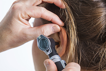
Ear specialists frequently use a surgical microscope to help diagnose and treat ear problems. The microscope provides intense light and magnification that help the doctor look down these small dark passages. The microscope also provides binocular vision which gives the doctor a sense of depth perception. This allows the doctor to treat the ears safely.
The doctor can see small areas inside the ears, which may help diagnose conditions of the ear. Doctors often perform surgery on the ear drum, middle ear bones, and mastoid using a microscope.
The tympanic membrane or ear drum is a thin piece of tissue that separates the ear canal from the middle ear space. Normally the tympanic membrane is an intact, complete sheet. Behind the tympanic membrane is a space called the middle ear. The middle ear naturally drains into the back of the nose through a passage called the Eustachian tube.
Bartolomeo Eustachio, an Italian anatomist who was famous for his meticulous dissections, discovered this passage that now bears his name. Improper Eustachian tube drainage can lead to middle ear infections or the accumulation of fluid in the middle ear.
ENT doctors are often called upon to help treat these problems. Since nasal problems may be a contributing factor, ENT doctors will usually carefully examine the nose as well. Also within the middle ear are the small bones of hearing. They are the malleus, incus, and stapes.
Their names come from their appearance. Malleus means hammer. Incus means anvil. Stapes means stirrup. Sound waves strike the eardrum, which then transmit vibrations to the middle ear bones which then vibrate and transmit the sound waves into the inner ear.
The sound waves vibrate the inner ear fluid and eventually move inner ear hair cells that convert the mechanical energy into electrical energy, which is then transmitted through the eighth cranial nerve to the brain. ENT doctors frequently examine patients’ ears and evaluate hearing problems.
There is also a balance organ in the inner ear that sends signals to the brain. Therefore, the inner ear can sometimes be a cause of balance problems or dizziness. Sometimes patients are sometimes referred to an ENT specialist to check whether a dizziness problem could be due to an ear problem.
Dr. James Go, MD, FACS
2290 West Eau Gallie Blvd
Suite 110
Melbourne, FL 32935
Phone: 321.421.7555
Fax: 321.421.7554
© 2023